Preprints
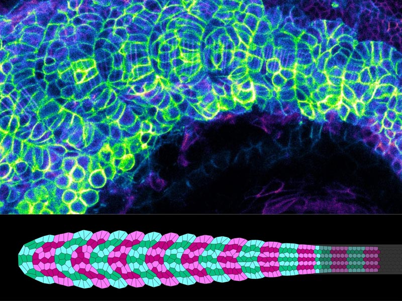
Loo YT, Chen J, Harrison R, Rito T, Theis S, Charras G, Briscoe J, and Saunders TE. Boundary constraints can determine pattern emergence.
Biorχiv
Athilingam T, Wilby EL, Bensidoun P, Trullo A, Verbrugghe M, Lagha M, Weil T, and Saunders TE. Regulation of bicoid mRNA throughout oogenesis and early embryogenesis impacts protein gradient formation.
Biorχiv
Amourda C, Chong J and Saunders TE. MicroRNAs buffer genetic variation at specific temperatures during embryonic development.
Biorχiv
2025
Le Y, Rajasekhar K, Loo TYJ, Saunders TE, Wohland T, and Winkler C. Midkine-a interacts with Ptprz1b to regulate neural plate convergence and midline formation in the developing zebrafish hindbrain. Developmental Biology; 521: 52-74. [PMID: 39924070]
Link
Wohland T, Saunders TE, and Chan CJ. Developmental biophysics (editorial). Biophysical Journal; 124, E1-E2. [PMID: 40015269]
Link
2024
Athilingam T, Nelanuthala AV, Breen C, Karedla N, Fritzsche M, Wohland T, and Saunders TE. Long-range formation of the Bicoid gradient requires multiple dynamic modes that spatially vary across the embryo. Development; 151: dev202128. [PMID: 38345326]
Link
Review: Wherley TJ, Thomas S, Millay DP, Saunders TE and Roy S. Molecular regulation of myocyte fusion. Current Topics in Developmental Biology; 158: 53-82 [PMID: 38670716]
Link
2023
Karkali K, Saunders TE, Panayotou G and Martín-Blanco E. JNK signaling in pioneer neurons organizes ventral nerve cord architecture in
Drosophila embryos. Nature Communications; 14: 675. [PMID: 36750572]
Link
2022
Mendieta-Serrano MA, Dhar S, Ng BH, Narayanan R, Lee JJY, Ong HT, Toh PJY, Röllin A, Roy S, and Saunders TE. Slow muscles guide fast myocyte fusion to ensure robust myotome formation despite the high spatiotemporal stochasticity of fusion events. Developmental cell; 57: 2095. [PMID: 36027918]
Link
Lai JKH, Toh PJY, Cognart HA, Chouhan G and Saunders TE. DNA-damage induced cell death in yap1; wwtr1 mutant epidermal basal cells. eLife; 11: e72302. [PMID: 35635436]
Link
Karkali K, Saunders TE, Vernon SW, Baines RA, Panayotou G, Martín-Blanco E. Condensation of the
Drosophila nerve cord is oscillatory and depends on coordinated mechanical interactions. Developmental Cell; 57: 867. [PMID: 35413236]
Link
Mahabaleshwar H, Asharani PV, Loo TYJ, Koh SY, Pitman MR, Kwok S, Ma J, Hu B, Lin F, Lok XL, Pitson SH, Saunders TE and Carney TJ. Slit-Robo Signalling Establishes a Sphingosine-1-Phosphate Gradient to Polarise Fin Mesenchyme and Establish Fin Morphology. EMBO Reports; 23: e54464. [PMID: 35679135]
Link
2021
Review: Saunders TE. The early
Drosophila embryo as a model system for quantitative biology. Cells and Development 2021;:203722. [PMID: 34298230]
Link
Yadav V, Tolwinski N, and Saunders TE. Spatiotemporal sensitivity of mesoderm specification to FGFR signalling in the
Drosophila embryo. Scientific Reports 2021; 11(1):14091. [PMID: 34238963]
Link
2020
Review: Mirth CK, Saunders TE, and Amourda C. Growing Up in a Changing World: Environmental Regulation of Development in Insects. Annual Reviews Entomology 2020;. [PMID: 32822557]
Link
Bosze B, Ono Y, Mattes B, Sinner C, Gourain V, Thumberger T, Tlili S, Wittbrodt J, Saunders TE, Strähle U, Schug A and Scholpp S. Pcdh18a regulates endocytosis of E-cadherin during axial mesoderm development in zebrafish. Histochemistry and Cell Biology 2020; 154: 463.
Link
Huang A, Rupprecht JF and Saunders TE. Embryonic geometry underlies phenotypic variation in decanalized conditions. eLife 2020: 9; e47380. [PMID: 32048988].
Link
Amourda C and Saunders TE. The mirtron miR-1010 functions in concert with its host gene SKIP to balance elevation of nAcRβ2. Scientific Reports 2020; 10:1. [PMID 32015391].
Link
Review: Huang A and Saunders TE. A matter of time: Formation and interpretation of the Bicoid morphogen gradient. Current Topics in Developmental Biology 2020; 137: 79. [PMID: 32143754]
Link
2019
Commentary: Saunders TE and Ingham PW. Open questions: how to get developmental biology into shape? BMC Biology 2019; 17:1. [PMID: 30795745]
Link
2018
Yin J, Lee R, Ono Y, Ingham PW and Saunders TE. Spatiotemporal coordination of FGF and Shh signaling underlies the specification of myoblasts in the zebrafish embryo. Developmental Cell 2018; 46; 73 [PMID: 30253169]
Link
Durrieu, L Kirrmaier D, Schneidt T, Kats I, Raghavan S, Hufnagel L, Saunders TE and Knop M. Bicoid gradient formation mechanism and dynamics revealed by protein lifetime analysis. Molecular Systems Biology 2018; 14: e8355 [PMID: 30181144]
Link
Chong J, Amourda C and Saunders TE. Temporal development of
Drosophila embryos is highly robust across a wide temperature range. Journal of the Royal Society Interface 2018; 15: 20180304. [PMID: 29997261]
Link
2017
Huang A, Amourda C, Zhang S, Tolwinski NS and Saunders TE. Decoding temporal interpretation of the morphogen Bicoid in the early
Drosophila embryo. eLife 2017; 6: e26258. [PMID: 28691901]
Link
Review: Amourda C and Saunders TE. Gene expression boundary scaling and organ size regulation in the
Drosophila embryo. Development, Growth and Differentiation 2017; 59: 21. [PMID: 28093727]
Link
2014
Pan KZ, Saunders TE, Flor-Parra I, Howard M and Chang F. Cortical regulation of cell size by a sizer cdr2p. eLife 2014; 3: e02040. [PMID:24642412]
Link
Erceg J, Saunders TE, Girardot C, Devos DP, Hufnagel L and Furlong EEM. Subtle changes in motif positioning cause tissue-specific effects on robustness of an enhancer's activity. PLoS Genetics 2014; 10: e1004060. [PMID: 24391522]
Link
2012
Saunders TE, Pan KZ, Angel A, Guan Y, Shah JV, Howard M and Chang F. Noise reduction in the intracellular pom1p gradient by a dynamic clustering mechanism. Developmental Cell 2012; 22: 558. [PMID: 22342545]
Link
2010
He F, Saunders TE, Wen Y, Cheung D, Jiao R, ten Wolde PR, Howard M and Ma J. Shaping a morphogen gradient for positional precision. Biophysical Journal 2010; 99: 69. [PMID: 20682246]
Link
Preprints
Yin J and Saunders TE. Shh induces symmetry breaking in the presomitic mesoderm by inducing tissue shear and orientated cell rearrangements
Biorχiv
2025
Theis S, Mendieta-Serrano MA, Chapa-y-Lazo B, Chen J, and Saunders TE. CellMet: Extracting 3D shape metrics from cells and tissues. PLoS Computational Biology in press.
Link
2024
de-Carvalho J, Tlili S, Saunders TE and Telley IA. The positioning mechanics of microtubule asters in Drosophila embryo explants. eLife; 12: RP90541. [PMID: 38426416]
Link
2023
Toh PJY, Sudol M and Saunders TE. Optogenetic control of YAP can enhance the rate of wound healing. Cellular and Molecular Biology Letters; 28: 39. [PMID: 37170209]
Link
2022
Toh PJY, Lai JKH, Hermann A, Destaing O, Sheets MP, Sudol M and Saunders TE. Optogenetic manipulation of YAP cellular localisation and function. EMBO Reports; 23: e54401. [PMID: 35876586]
Link
de-Carvalho J, Tlili S, Hufnagel L, Saunders TE and Telley IA. Aster repulsion drives local ordering in an active system. Development 2022; 149: dev199997. [PMID: 35001104]
Link
2021
Tiwari P, Rengarajan H, and Saunders TE. Scaling of Internal Organs during Drosophila Embryonic Development. Biophysical Journal 2021; 120: 1-13. [PMID: 34087212]
Link
Review: Narayanan R, Mendieta-Serrano MA, and Saunders TE. The role of cellular active stresses in shaping the zebrafish body axis. Current Opinion in Cell Biology 2021; 73:69-77. [PMID: 34303916]
Link
Review: Athilingam T, Tiwari P, Toyama Y, and Saunders TE. Mechanics of epidermal morphogenesis in the Drosophila pupa. Seminars in Cell and Developmental Biology 2021; [PMID: 34167884]
Link
Review: Zhang S and Saunders TE. Mechanical processes underlying precise and robust cell matching. Seminars in Cell and Developmental Biology.
Link
Commentary: Lenne PF
el al. Roadmap on multiscale coupling of biochemical and mechanical signals during development. Physical Biology 2020;. [PMID: 33276350]
Link
2020
Colin L, Chevallier A, Tsugawa S, Gacon F, Godin C, Viasnoff V, Saunders TE, and Hamant O. Cortical tension overrides geometrical cues to orient microtubules in confined protoplasts. PNAS 2020;. [PMID: 33288703]
Link
Zhang S, Teng X, Toyama Y, and Saunders TE. Periodic Oscillations of Myosin-II Mechanically Proofread Cell-Cell Connections to Ensure Robust Formation of the Cardiac Vessel. Current Biology 2020; 30: 3364. [PMID: 32679105]
Link
Review: Hamant O, and Saunders TE. Shaping Organs: Shared Structural Principles Across Kingdoms. Annual Reviews Cell and Developmental Biology 2020;. [PMID: 32628862]
Link
2019
Tlili S, Yin J, Rupprecht J-F, Mendieta-Serrano MA, Weissbart G, Verma N, Teng X, Toyama Y, Prost J and Saunders TE. Shaping the zebrafish myotome by intertissue friction and active stress. PNAS 2019; 116: 25430. [PMID: 31772022]
Link
2018
Zhang S, Amourda C, Garfield D and Saunders TE. Selective filopodia adhesion ensures robust cell matching in the Drosophila heart. Developmental Cell 2018; 46: 189. [PMID: 30016621]
Link
2017
Sun Z, Amourda C, Shagirov M, Hara Y, Saunders TE and Toyama Y. Basolateral protrusion and apical contraction cooperatively drive Drosophila germ-band extension. Nature Cell Biology 2017; 19:375. [PMID: 28346438]
Link
Review: Saunders TE. Imag(in)ing growth and form. Mechanisms of Development 2017; 145: 13. [PMID: 28351699]
Link
2015
Rauzi M, Krzic U, Saunders TE, Krajnc M, Ziherl P, Hufnagel L and Leptin M. Embryo-scale tissue mechanics during
Drosophila gastrulation movements. Nature Communications 2015; 6: 1-12. [PMID: 26497898]
Link
Preprints
Lou Y, Theis S, Rupprecht JF, Saunders TE, and Hiraiwa T. Stress anisotropy in 3D active curved structures.
Biorχiv
Loo TYJ, Mahabaleshwar H, Carney T and Saunders TE. Echolocation-like model of directed cell migration within growing tissues.
Biorχiv
2025
Commentary: Saunders TE. News and Views: Topology Drives Cell Fate. Nature Physics; 21, 508-509.
Link
Zhu S, Loo YT, Veerapathiran S, Loo TYJ, Tran BN Teh C, Zhong J, Saunders TE, and Wohland T. Receptor binding and tortuosity explain morphogen local-to-global diffusion coefficient transition. Biophysical Journal; 124, 963-979. [PMID: 39049492]
Link
Tlili S, Shagirov M, Zhang S and Saunders TE. Interfacial energy constraints are sufficient to align cells over large distances. Biophysical Journal; 124, 1011-1023. [PMID: 40081366]
Link
2023
Lou Y, Rupprecht JF, Theis S, Hiraiwi T and Saunders TE. Curvature-induced cell rearrangements in biological tissues. Physical Review Letters 2023; 130: 108401. [PMID: 36962052]
Link
2020
Thamm A, Saunders TE and Dolan L. MpFEW RHIZOIDS1 miRNA-mediated lateral inhibition controls rhizoid cell patterning in Marchantia polymorpha. Current Biology 2020; 30: 1905. [PMID: 32243863]
Link
Bezeljak U, Loya H, Kaczmarek B, Saunders TE, Loose M. Stochastic activation and bistability in a Rab GTPase regulatory network. PNAS 2020; 117: 6540. [PMID: 32161136]
Link
2019
Connahs H, Tlili S, van Creij J, Loo TYJ, Banerjee TD, Saunders TE and Monteiro A. Activation of butterfly eyespots by Distal-less is consistent with a reaction-diffusion process. Development 2019; 146: dev169367 [PMID: 30992277]
Link
2017
Kaur P, Saunders TE and Tolwinski NS. Coupling optogenetics and light-sheet microscopy, a method to study Wnt signaling during embryogenesis. Scientific Reports 2017; 7: 1-11. [PMID: 29192250]
Link
Singh AP, Galland R, Finch-Edmondson ML, Grenci G, Sibarita J-B, Studer V, Viasnoff V and Saunders TE. 3D protein dynamics in the cell nucleus. Biophysical Journal (2017); 112: 133. [PMID: 28076804]
Link
2015
Krieger JW, Singh AP, Bag N, Garbe CS, Saunders TE, Langowski J and Wohland T. Imaging fluorescence (cross-) correlation spectroscopy in live cells and organisms. Nature Protocols 2015; 10: 1948. [PMID: 26540588]
Link
Richards DM and Saunders TE. Spatiotemporal analysis of different mechanisms for interpreting morphogen gradients. Biophysical Journal 2015; 108: 2061-2073. [PMID: 25902445]
Link
Saunders TE. Aggregation-fragmentation model of robust concentration gradient formation. Physical Review E 2015; 91: 022704. [PMID: 25768528]
Link
2012
Krzic U, Gunther S, Saunders TE, Streichan SJ and Hufnagel L. Multiview light-sheet microscope for rapid
in toto imaging. Nature Methods 2012; 9: 730. [PMID: 22660739]
Link
2010
Andreanov A, Chalker JT, Saunders TE and Sherrington D. Spin-glass transition in geometrically frustrated antiferromagnets with weak disorder. Physical Review B 2010; 81: 014406
Link
2009
Saunders TE and Howard M. When it pays to rush: interpreting morphogen gradients prior to steady-state. Physical Biology 2009; 6: 046020. [PMID: 19940351]
Link
Saunders TE and Howard M. Morphogen profiles can be optimized to buffer against noise. Physical Review E 2009; 80: 041902. [PMID: 19905337]
Link
2008
Pickles T, Saunders TE and Chalker JT. Critical phenomena in a highly constrained classical spin system: Néel ordering from the Coulomb phase. Europhysics Letters 2008; 84: 36002
Link
Saunders TE and Chalker JT. Structural phase transitions in geometrically frustrated antiferromagnets. Physical Review B 2008; 77: 214438
Link
2007
Saunders TE and Chalker JT. Spin freezing in geometrically frustrated antiferromagnets with weak disorder. Physical Review Letters 2007; 98: 157201
Link
and PLoS Complex Systems.
I recently co-edited a special issue for Biophyiscal Journal on Developmental Biophysics with Thorsten Wohland and Chii Jou Chan. I have edited a special issue for
on mechanobiology in development. I also edited a special issue for Mechanisms of Development (now Cells and Development) with Prof. Philip Ingham on 100 years since "On Growth and Form" by D'Arcy Wentworth-Thompson:
I have reviewed for a large range of journals, including: Nature, Cell, Nature Cell Biology, Current Biology, PNAS, Development, Developmental Cell, PLoS Biology, PLoS Computational Biology, Molecular Systems Biology, Physical Review Letters, Physical Review E, Cells and Development, Molecular Biology of the Cell, EMBO, Cell Systems, Cell Reports.
I have reviewed for a broad range of international grant agencies, including: BBSRC (UK), Wellcome (UK), DFG (Germany), ERC (EU) and Agence Nationale de la Recherche (France).
